


Left heart catheterization
Catheterization - left heartLeft heart catheterization is the passage of a thin flexible tube (catheter) into the left side of the heart. It is done to diagnose or treat certain heart problems.
How the Test is Performed
You may be given a mild medicine (sedative) before the procedure starts. The medicine is to help you relax. The health care provider will place an IV into your arm to give medicines. You will lie on a padded table. Your doctor (most often a heart specialist called a cardiologist) will make a small puncture on your body near one of your arteries. A flexible tube (catheter) is inserted into and threaded through the artery. It will be placed in your wrist, arm or your upper leg (groin). You will most likely be awake during the procedure.
Live x-ray pictures are used to help guide the catheters up into your heart and arteries. Dye (sometimes called "contrast") will be injected into your body. This dye will highlight blood flow through the arteries. This helps show blockages in the blood vessels that lead to your heart.
The catheter is then moved through the aortic valve into the left side of your heart. The pressure is measured in the heart in this position. Other procedures can also be done at this time, such as:
- Ventriculography to check the heart's pumping function.
Ventriculography
Left heart ventricular angiography is a procedure to look at the left-sided heart chambers and the function of the left-sided valves. It is sometime...
 ImageRead Article Now Book Mark Article
 - Coronary angiography to look at the coronary arteries.
Coronary angiography
Coronary angiography is a procedure that uses a special dye (contrast material) and x-rays to see how blood flows through the arteries in your heart....
 ImageRead Article Now Book Mark Article
 - Angioplasty, with or without stenting, to correct blockages in the arteries is then performed.
Angioplasty
Angioplasty is a procedure to open narrowed or blocked blood vessels that supply blood to the heart. These blood vessels are called the coronary art...
 ImageRead Article Now Book Mark Article
The procedure may last from less than 1 hour to several hours.
How to Prepare for the Test
In most cases, you should not eat or drink for 8 hours before the test. (your cardiologist may give you different directions.)
The procedure will take place in the hospital. You may be admitted the night before the test, but it is more common to come to the hospital the morning of the procedure. In some cases, this procedure is done after you have already been admitted to the hospital possibly on an emergency basis.
You will wear a hospital gown. Your cardiologist will explain the procedure and its risks. You must sign a consent form.
How the Test will Feel
The sedative will help you relax before the procedure. However, you will be awake and able to follow instructions during the test.
You will be given local numbing medicine (anesthesia) before the catheter is inserted. You will feel some pressure as the catheter is inserted. However, you should not feel any pain. You may have some discomfort from lying still for a long period of time.
Why the Test is Performed
The procedure is done to look for:
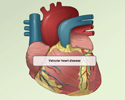
- Cardiac valve disease
- Cardiac tumors
- Heart defects (such as ventricular septal defects)
Ventricular septal defects
A ventricular septal defect is a hole in the wall that separates the right and left ventricles of the heart. Ventricular septal defect is one of the...
 ImageRead Article Now Book Mark Article
 - Problems with heart function
The procedure may also be done to evaluate and possibly repair certain types of heart defects, or to open a narrowed heart valve.
When this procedure is done with coronary angiography to examine the arteries that feed the heart muscle, it can be done with angioplasty to open blocked arteries or bypass grafts. This can be because of a heart attack or angina.
Coronary angiography
Coronary angiography is a procedure that uses a special dye (contrast material) and x-rays to see how blood flows through the arteries in your heart....
Angina
Angina is a type of chest discomfort or pain due to poor blood flow through the blood vessels (coronary arteries) of the heart muscle (myocardium). ...

The procedure can also be used to:
- Collect blood samples from the heart
- Determine pressure and blood flow in the heart's chambers
- Take x-ray pictures of the left ventricle (main pumping chamber) of the heart (ventriculography)
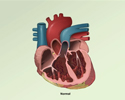
Normal Results
A normal result means the heart is normal in:
- Size
- Motion
- Thickness
- Pressure
The normal result also means arteries are normal.
What Abnormal Results Mean
Abnormal results may be a sign of cardiac disease or heart defects, including:
- Aortic insufficiency
Aortic insufficiency
Aortic regurgitation is a heart valve disease in which the aortic valve does not close tightly. This allows blood to flow from the aorta (the larges...
 ImageRead Article Now Book Mark Article
 - Aortic stenosis
Aortic stenosis
The aorta is the main artery that carries blood out of the heart to the rest of the body. Blood flows out of the heart and into the aorta through th...
 ImageRead Article Now Book Mark Article
 - Coronary artery disease
Coronary artery disease
Stable angina is chest pain or discomfort that most often occurs with activity or emotional stress. Angina is due to poor blood flow through the blo...
 ImageRead Article Now Book Mark Article
 - Heart enlargement
Heart enlargement
Cardiomyopathy is disease in which the heart muscle becomes weakened, stretched, or has another structural problem. Dilated cardiomyopathy is a condi...
 ImageRead Article Now Book Mark Article
 - Mitral regurgitation
Mitral regurgitation
Mitral regurgitation is a disorder in which the mitral valve on the left side of the heart does not close properly. Regurgitation means leaking from ...
ImageRead Article Now Book Mark Article - Mitral stenosis
Mitral stenosis
Mitral stenosis is a disorder in which the mitral valve does not fully open. This restricts the flow of blood.
ImageRead Article Now Book Mark Article - Ventricular aneurysms
Aneurysms
An aneurysm is an abnormal widening or ballooning of a part of an artery due to weakness in the wall of the blood vessel.
 ImageRead Article Now Book Mark Article
 - Atrial septal defect
Atrial septal defect
Atrial septal defect (ASD) is a heart defect that is present at birth (congenital). As a baby develops in the womb, a wall (septum) forms that divide...
 ImageRead Article Now Book Mark Article
 - Ventricular septal defect
Ventricular septal defect
A ventricular septal defect is a hole in the wall that separates the right and left ventricles of the heart. Ventricular septal defect is one of the...
ImageRead Article Now Book Mark Article - Heart failure
- Cardiomyopathy
Cardiomyopathy
Cardiomyopathy is disease in which the heart muscle becomes weakened, stretched, or has another structural problem. It often contributes to the hear...
ImageRead Article Now Book Mark Article
Risks
Complications may include:
- Cardiac arrhythmias
Arrhythmias
An arrhythmia is a disorder of the heart rate (pulse) or heart rhythm. The heart can beat too fast (tachycardia), too slow (bradycardia), or irregul...
ImageRead Article Now Book Mark Article - Cardiac tamponade
Cardiac tamponade
Cardiac tamponade is pressure on the heart that occurs when blood or fluid builds up in the space between the heart muscle and the outer covering sac...
ImageRead Article Now Book Mark Article - Embolism from blood clots at the tip of the catheter to the brain or other organs
Embolism
Blood clots are clumps that occur when blood hardens from a liquid to a solid. A blood clot that forms inside one of your veins or arteries is calle...
 ImageRead Article Now Book Mark Article
 - Heart attack
Heart attack
Most heart attacks are caused by a blood clot that blocks one of the coronary arteries. The coronary arteries bring blood and oxygen to the heart. ...
 ImageRead Article Now Book Mark Article
 - Injury to the artery
- Infection
- Kidney damage from contrast (dye)
- Low blood pressure
- Reaction to the contrast material
- Stroke
Stroke
A stroke occurs when blood flow to a part of the brain stops. A stroke is sometimes called a "brain attack. " If blood flow is cut off for longer th...
 ImageRead Article Now Book Mark Article
References
Dengas GD, Mehran R. Coronary angiography and intravascular imaging. In: Libby P, Bonow RO, Mann DL, Tomaselli GF, Bhatt DL, Solomon SD, eds. Braunwald's Heart Disease: A Textbook of Cardiovascular Medicine. 12th ed. Philadelphia, PA: Elsevier; 2022:chap 21.
Kern MJ, Seto AH, Hermann J. Invasive hemodynamic diagnosis of cardiac disease. In: Libby P, Bonow RO, Mann DL, Tomaselli GF, Bhatt DL, Solomon SD, eds. Braunwald's Heart Disease: A Textbook of Cardiovascular Medicine. 12th ed. Philadelphia, PA: Elsevier; 2022:chap 22.
Lawton J, Tamis-Holland, J. et al. 2021 ACC/AHA/SCAI Guideline for coronary artery revascularization: A report of the American College of Cardiology/American Heart Association Joint Committee on Clinical Practice Guidelines. J Am Coll Cardiol. 2022;79(2):e21–e129. PMID: 34895950 pubmed.ncbi.nlm.nih.gov/34895950/.
Left heart catheterization - illustration
Left heart catheterization involves the passage of a catheter (a thin flexible tube) into the left side of the heart to obtain diagnostic information about the left side of the heart or to provide therapeutic interventions in certain types of heart conditions. The test can determine pressure and blood flow in the heart's chambers, collect blood samples from the heart, and examine the arteries of the heart by X-ray (fluoroscopy).
Left heart catheterization
illustration
Review Date: 7/14/2024
Reviewed By: Michael A. Chen, MD, PhD, Associate Professor of Medicine, Division of Cardiology, Harborview Medical Center, University of Washington Medical School, Seattle, WA. Also reviewed by David C. Dugdale, MD, Medical Director, Brenda Conaway, Editorial Director, and the A.D.A.M. Editorial team.
 All rights reserved.