


Electrocardiogram
ECG; EKGAn electrocardiogram (ECG) is a test that records the electrical activity of the heart.
How the Test is Performed
You will be asked to lie down. A technician will clean several areas on your arms, legs, and chest, and then will attach small patches called electrodes to those areas. It may be necessary to shave or clip some hair so the patches stick to the skin. The number of patches used may vary.
The patches are connected by wires to a machine that turns the heart's electrical signals into wavy lines, which are often printed on paper. The health care provider reviews the test results.
You will need to remain still during the procedure. The technician may also ask you to hold your breath for a few seconds as the test is being done.
It is important to be relaxed and warm during an ECG recording because any movement, including shivering, can alter the results.
Sometimes this test is done while you are exercising or under light stress to look for changes in the heart. This type of ECG is often called a stress test.
How to Prepare for the Test
Make sure your provider knows about all the medicines you are taking. Some medicines can interfere with test results.
Do not exercise or drink cold water immediately before an ECG because these actions may cause false results.
How the Test will Feel
An ECG is painless. No electricity is sent through the body. The electrodes may feel cold when first applied. In rare cases, some people may develop a rash or irritation where the patches were placed.
Why the Test is Performed
An ECG is used to measure:
- Any damage to your heart
- How fast your heart is beating and whether it is beating normally
- The effects of medicines or devices used to control your heart (such as a pacemaker)
Pacemaker
A pacemaker is a small, battery-operated device. This device senses when your heart is beating too slowly. It sends a signal to your heart that mak...
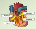
Read Article Now Book Mark Article - The size and position of your heart chambers
An ECG is often the first test done to determine whether a person has heart disease. Your provider may order this test if:
Heart disease
Coronary heart disease is a narrowing of the blood vessels that supply blood and oxygen to the heart. Coronary heart disease (CHD) is also called co...
- You have chest pain or palpitations
Chest pain
Chest pain is discomfort or pain that you feel anywhere along the front of your body between your neck and upper abdomen.
 ImageRead Article Now Book Mark Article
Palpitations
Palpitations are feelings or sensations that your heart is pounding or racing. They can be felt in your chest, throat, or neck. You may:Have an unpl...
 ImageRead Article Now Book Mark Article
 - You are scheduled for surgery
- You have had heart problems in the past
- You have a strong history of heart disease in the family
Normal Results
Normal test results most often include:
- Heart rate: 60 to 100 beats per minute
- Heart rhythm: Consistent and even
What Abnormal Results Mean
Abnormal ECG results may be a sign of:
- Damage or changes to the heart muscle
- Changes in the amount of the electrolytes (such as potassium and calcium) in the blood
- Congenital heart defect
- Enlargement of the heart
Enlargement of the heart
Cardiomyopathy is disease in which the heart muscle becomes weakened, stretched, or has another structural problem. Dilated cardiomyopathy is a condi...
 ImageRead Article Now Book Mark Article
 - Fluid or swelling in the sac around the heart
- Inflammation of the heart (myocarditis)
Inflammation of the heart
Pericarditis is a condition in which the sac-like covering around the heart (pericardium) becomes inflamed.
 ImageRead Article Now Book Mark Article
 - Past or current heart attack
Heart attack
Most heart attacks are caused by a blood clot that blocks one of the coronary arteries. The coronary arteries bring blood and oxygen to the heart. ...
 ImageRead Article Now Book Mark Article
 - Poor blood supply to the heart arteries
- Abnormal heart rhythms (arrhythmias)
Arrhythmias
An arrhythmia is a disorder of the heart rate (pulse) or heart rhythm. The heart can beat too fast (tachycardia), too slow (bradycardia), or irregul...
 ImageRead Article Now Book Mark Article
Some heart problems that can lead to changes on an ECG test include:
- Atrial fibrillation/flutter
Atrial fibrillation/flutter
Atrial fibrillation (AFib) and atrial flutter are common types of abnormal heart rhythms (arrhythmias) which affect the upper chambers (atria) of the...
 ImageRead Article Now Book Mark Article
 - Heart attack
- Heart failure
Heart failure
Heart failure is a condition in which the heart is no longer able to pump oxygen-rich blood to the rest of the body efficiently. This causes symptom...
ImageRead Article Now Book Mark Article - Multifocal atrial tachycardia
Multifocal atrial tachycardia
Multifocal atrial tachycardia (MAT) is a rapid heart rate. It occurs when too many signals (electrical impulses) are sent from the upper heart (atri...
ImageRead Article Now Book Mark Article - Paroxysmal supraventricular tachycardia
Paroxysmal supraventricular tachycardia
Paroxysmal supraventricular tachycardia (PSVT) is episodes of a rapid heart rate that start in a part of the heart above the ventricles. "Paroxysmal...
 ImageRead Article Now Book Mark Article
 - Sick sinus syndrome
Sick sinus syndrome
Normally, the heartbeat starts in an area in the top chambers of the heart (atria). This area is the heart's pacemaker. It is called the sinoatrial...
 ImageRead Article Now Book Mark Article
 - Wolff-Parkinson-White syndrome
Wolff-Parkinson-White syndrome
Wolff-Parkinson-White (WPW) syndrome is a condition in which there is an extra electrical pathway in the heart that leads to periods of rapid heart r...
 ImageRead Article Now Book Mark Article
Risks
There are no risks.
Considerations
The accuracy of the ECG depends on the condition being tested. A heart problem may not always show up on the ECG. Some heart conditions never produce any specific ECG changes.
References
Brady WJ, Harrigan RA, Chan TC. Basic electrocardiographic techniques. In: Roberts JR, Custalow CB, Thomsen TW, eds. Roberts and Hedges' Clinical Procedures in Emergency Medicine and Acute Care. 7th ed. Philadelphia, PA: Elsevier; 2019:chap 14.
Ganz L, Link MS. Electrocardiography. In: Goldman L, Cooney KA, eds. Goldman-Cecil Medicine. 27th ed. Philadelphia, PA: Elsevier; 2024:chap 42.
Mirvis DM, Goldberger AL. Electrocardiography. In: Libby P, Bonow RO, Mann DL, Tomaselli GF, Bhatt DL, Solomon SD, eds. Braunwald's Heart Disease: A Textbook of Cardiovascular Medicine. 12th ed. Philadelphia, PA: Elsevier; 2022:chap 14.
ECG - illustration
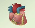
The electrocardiogram (ECG) is used extensively in the diagnosis of heart disease, from congenital heart disease in infants to myocardial infarction and myocarditis in adults. Several different types of electrocardiogram exist.
ECG
illustration
Atrioventricular block - ECG tracing - illustration
This picture shows an ECG (electrocardiogram, EKG) of a person with an abnormal rhythm (arrhythmia) called an atrioventricular (AV) block. P waves show that the top of the heart received electrical activity. Each P wave is usually followed by the tall (QRS) waves. QRS waves reflect the electrical activity that causes the heart to contract. When a P wave is present and not followed by a QRS wave (and heart contraction), there is an atrioventricular block, and a very slow pulse (bradycardia).
Atrioventricular block - ECG tracing
illustration
High blood pressure tests - illustration
Routine lab tests are recommended before beginning treatment of high blood pressure to determine organ or tissue damage or other risk factors. These lab tests include urinalysis, blood cell count, blood chemistry (potassium, sodium, creatinine, fasting glucose, total cholesterol and HDL cholesterol), and an ECG (electrocardiogram). Additional tests may be recommended based on your condition.
High blood pressure tests
illustration
Electrocardiogram (ECG) - illustration
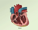
An electrocardiogram is a test that measures the electrical activity of the heart. This includes the rate and regularity of beats as well as the size and position of the chambers, any damage to the heart, and effects of drugs or devices to regulate the heart.
Electrocardiogram (ECG)
illustration
ECG electrode placement - illustration
An ECG is very useful in determining whether a person has heart disease. During an ECG electrodes are affixed to each arm and leg and to the chest.
ECG electrode placement
illustration


ECG - illustration
The electrocardiogram (ECG) is used extensively in the diagnosis of heart disease, from congenital heart disease in infants to myocardial infarction and myocarditis in adults. Several different types of electrocardiogram exist.
ECG
illustration
Atrioventricular block - ECG tracing - illustration
This picture shows an ECG (electrocardiogram, EKG) of a person with an abnormal rhythm (arrhythmia) called an atrioventricular (AV) block. P waves show that the top of the heart received electrical activity. Each P wave is usually followed by the tall (QRS) waves. QRS waves reflect the electrical activity that causes the heart to contract. When a P wave is present and not followed by a QRS wave (and heart contraction), there is an atrioventricular block, and a very slow pulse (bradycardia).
Atrioventricular block - ECG tracing
illustration
High blood pressure tests - illustration
Routine lab tests are recommended before beginning treatment of high blood pressure to determine organ or tissue damage or other risk factors. These lab tests include urinalysis, blood cell count, blood chemistry (potassium, sodium, creatinine, fasting glucose, total cholesterol and HDL cholesterol), and an ECG (electrocardiogram). Additional tests may be recommended based on your condition.
High blood pressure tests
illustration
Electrocardiogram (ECG) - illustration
An electrocardiogram is a test that measures the electrical activity of the heart. This includes the rate and regularity of beats as well as the size and position of the chambers, any damage to the heart, and effects of drugs or devices to regulate the heart.
Electrocardiogram (ECG)
illustration
ECG electrode placement - illustration
An ECG is very useful in determining whether a person has heart disease. During an ECG electrodes are affixed to each arm and leg and to the chest.
ECG electrode placement
illustration
Review Date: 5/8/2024
Reviewed By: Thomas S. Metkus, MD, Assistant Professor of Medicine and Surgery, Johns Hopkins University School of Medicine, Baltimore, MD. Also reviewed by David C. Dugdale, MD, Medical Director, Brenda Conaway, Editorial Director, and the A.D.A.M. Editorial team.
© 1997- A.D.A.M., a business unit of Ebix, Inc. Any duplication or distribution of the information contained herein is strictly prohibited.
 All rights reserved.