


Neuroretinitis
Neuroretinitis is inflammation of the retina and optic nerve of the eye. The condition can be caused by bacteria, viruses or autoimmune disease. It shares some features of optic neuritis. There is mostly central visual loss, which often recovers after some months, but often not completely.
Retina
The retina is the light-sensitive layer of tissue at the back of the eyeball. Images that come through the eye's lens are focused on the retina. Th...

Optic neuritis
The optic nerve carries images of what the eye sees to the brain. When this nerve become swollen or inflamed, it is called optic neuritis. It may c...

There is no treatment that has proven to be helpful.
References
Cioffi GA, Liebmann JM. Bartonella infections. In: Goldman L, Cooney KA, eds. Goldman-Cecil Medicine. 27th ed. Philadelphia, PA: Elsevier; 2024:chap 291.
Moss HE, Guercio JR, Balcer LJ. Inflammatory optic neuropathies and neuroretinitis. In: Yanoff M, Duker JS, eds. Ophthalmology. 6th ed. Philadelphia, PA: Elsevier; 2023:chap 9.7.
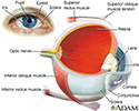
Eye - illustration
The eye is the organ of sight, a nearly spherical hollow globe filled with fluids (humors). The outer layer (sclera, or white of the eye, and cornea) is fibrous and protective. The middle layer (choroid, ciliary body and the iris) is vascular. The innermost layer (retina) is sensory nerve tissue that is light sensitive. The fluids in the eye are divided by the lens into the vitreous humor (behind the lens) and the aqueous humor (in front of the lens). The lens itself is flexible and suspended by ligaments which allow it to change shape to focus light on the retina, which is composed of sensory neurons.
Eye
illustration
Retina - illustration
The retina is the internal layer of the eye that receives and transmits focused images. The retina is normally red due to its rich blood supply.
Retina
illustration
Review Date: 11/8/2023
Reviewed By: Franklin W. Lusby, MD, Ophthalmologist, Lusby Vision Institute, La Jolla, CA. Also reviewed by David C. Dugdale, MD, Medical Director, Brenda Conaway, Editorial Director, and the A.D.A.M. Editorial team.
 All rights reserved.