


Basal cell skin cancer
Basal cell carcinoma; Rodent ulcer; Skin cancer - basal cell; Cancer - skin - basal cell; Nonmelanoma skin cancer; Basal cell NMSC; Basal cell epitheliomaBasal cell cancer is the most common form of cancer in the United States. Most skin cancers are basal cell cancer.
Other common types of skin cancer are:
- Squamous cell cancer
Squamous cell cancer
Squamous cell skin cancer is the second most common type of cancer in the United States. Other common types of skin cancer are:Basal cell cancerMelan...
 ImageRead Article Now Book Mark Article
 - Melanoma
Melanoma
Melanoma is the most dangerous type of skin cancer. It is also the rarest. It is the leading cause of death from skin disease. Other common types o...
 ImageRead Article Now Book Mark Article
Causes
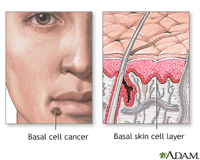
The top layer of the skin is called the epidermis. The bottom layer of the epidermis is the basal cell layer. With basal cell cancer, cells in this layer are the ones that become cancerous. Most basal cell cancers occur on skin that is regularly exposed to sunlight or other ultraviolet radiation.
This type of skin cancer is most common in people over age 50. But it can also occur in younger people who have had extensive sun exposure. Basal cell cancer is almost always slow-growing. It rarely spreads to other parts of the body.
You are more likely to develop basal cell cancer if you have:
- Light-colored or freckled skin
- Blue, green, or grey eyes
- Blond or red hair
- Overexposure to x-rays or other forms of radiation
- Many moles
- Close relatives who have or had skin cancer
- Many severe sunburns early in life
- Long-term daily sun exposure (such as the sun exposure received by people who work outside)
Other risk factors include:
- Smoking
- Weakened immune system, such as being on medicines that suppress the immune system after an organ transplant
- Inherited skin diseases, such as nevoid basal cell carcinoma syndrome
Nevoid basal cell carcinoma syndrome
Nevoid basal cell carcinoma syndrome is a group of conditions passed down through families. The disorder involves the skin, nervous system, eyes, en...
 ImageRead Article Now Book Mark Article
 - Having had photodynamic therapy
Photodynamic therapy
Photodynamic therapy (PDT) uses a medicine together with a special type of light to kill cancer cells.
Read Article Now Book Mark Article
Symptoms
Basal cell cancer usually grows slowly and is often painless. It may not look that different from your normal skin. You may have a skin bump or growth that is:
- Pearly or waxy
- White or light pink
- Flesh-colored or brown
- A red, scaly patch of skin
In some cases, the skin is just slightly raised, or even flat.
You may have:
- A skin sore that bleeds easily
- A sore that does not heal
- Oozing or crusting spots in a sore
- A scar-like sore without having injured the area
- Irregular blood vessels in or around the spot
- A sore with a depressed (sunken) area in the middle
Exams and Tests
Your health care provider will check your skin and look at the size, shape, color, and texture of any suspicious areas.
If your provider thinks you might have skin cancer, a piece of skin will be removed. This is called a skin biopsy. The sample is sent to a lab for examination under a microscope.
Skin biopsy
A skin lesion biopsy is when a small amount of skin is removed so it can be examined under a microscope. The skin is tested to look for skin conditi...

A skin biopsy must be done to confirm basal cell cancer or other skin cancers. The diagnosis cannot be based only on the appearance of your skin.
Treatment
The treatment depends on the size, depth, and location of the skin cancer and your overall health. Each treatment has its risks and benefits. You and your provider can discuss the treatment that's right for you.
Treatment may involve any of the following:
- Excision: Cutting out the skin cancer and stitching the skin together
- Curettage and electrodessication: Scraping away cancer cells and using electricity to kill any that remain; used to treat cancers that are not large or deep; often curettage is used alone without electrodessication
- Cryosurgery: Freezing the cancer cells, which kills them; used to treat cancers that are not large or deep
Cryosurgery
Cryotherapy is a method of superfreezing tissue in order to destroy it. This article discusses cryotherapy of the skin.
Read Article Now Book Mark Article - Medicine: Skin creams containing imiquimod or 5-fluorouracil for cancers that are not large or deep
- Mohs surgery: Removing a layer of skin and looking at it immediately under a microscope, then removing additional layers of skin until there are no signs of the cancer; usually used for skin cancers on the nose, ears, and other areas of the face
Mohs surgery
Mohs micrographic surgery is a way to treat and cure certain skin cancers. Surgeons trained in the Mohs procedure can do this surgery. It allows sk...
 ImageRead Article Now Book Mark Article
 - Photodynamic therapy: Using a light-activated chemical to treat cancers that are not large or deep
- Radiation therapy: May be used if a basal cell cancer cannot be treated with surgery
Radiation therapy
Radiation therapy uses high-powered radiation (such as x-rays or gamma rays), particles, or radioactive seeds to kill cancer cells.
 ImageRead Article Now Book Mark Article
 - Chemotherapy: May be used in the rare instances of basal cell cancer that has spread to other parts of the body or that cannot be treated with surgery
Chemotherapy
The term chemotherapy is used to describe cancer-killing drugs. Chemotherapy may be used to:Cure the cancerShrink the cancerPrevent the cancer from ...
 ImageRead Article Now Book Mark Article
 - Biologic therapies (immunotherapies): Medicines that target and kill basal cell skin cancer and are used when standard treatments do not work
Immunotherapies
Immunotherapy is a type of cancer treatment that relies on the body's infection-fighting system (immune system). It uses substances made by the body...
Read Article Now Book Mark Article
Support Groups
You can ease the stress of illness by joining a cancer support group. Sharing with others who have common experiences and problems can help you not feel alone.
Cancer support group
The following organizations are good resources for information on cancer:American Cancer Society. Support and online communities. www. cancer. org/...
Outlook (Prognosis)
Most of these cancers are cured when treated early. Some basal cell cancers return in the same location. Smaller ones are less likely to come back.
Basal cell skin cancer almost never spreads beyond the original location. Left untreated, however, it may spread into surrounding areas and nearby tissues and bone.
When to Contact a Medical Professional
Contact your provider for an appointment if you have a sore or spot on your skin that changes in:
- Appearance
- Color
- Size
- Texture
Also contact your provider if a spot becomes painful or swollen, or if it starts to bleed or itch.
Prevention
Experts disagree about the value of routine skin exams by your provider. But all agree it's important to know your own skin and to contact your provider if you notice anything unusual.
The best way to prevent skin cancer is to reduce your exposure to sunlight. Always use sunscreen:
Reduce your exposure to sunlight
Many skin changes, such as skin cancer, wrinkles, and age spots are caused by exposure to the sun. This is because the damage caused by the sun is p...

- Apply sunscreen with sun protection factor (SPF) of at least 30, even when you are going outdoors for a short time.
- Apply a large amount of sunscreen on all exposed areas, including ears and feet.
- Look for sunscreen that blocks both UVA and UVB light.
- Use a water-resistant sunscreen.
- Apply sunscreen at least 30 minutes before going out. Follow package instructions about how often to reapply. Be sure to reapply after swimming or sweating.
- Use sunscreen in winter and on cloudy days too.
Other measures to help you avoid too much sun exposure:
- Ultraviolet light is most intense between 10 a.m. and 4 p.m. Try to avoid the sun during these hours.
- Protect your skin by wearing wide-brim hats, long-sleeve shirts, long skirts, or pants. You can also buy sun-protective clothing.
- Avoid surfaces that reflect light more, such as water, sand, concrete, and areas that are painted white.
- The higher the altitude, the faster your skin burns.
- Do not use sun lamps and tanning beds (salons). Spending 15 to 20 minutes at a tanning salon is as dangerous as a day spent in the sun.
References
American Cancer Society website. How to do a skin self-exam. www.cancer.org/cancer/risk-prevention/sun-and-uv/skin-exams.html. Updated June 26, 2024. Accessed August 6, 2025.
Dinulos JGH. Premalignant and malignant nonmelanoma skin tumors. In: Dinulos JGH, ed. Habif's Clinical Dermatology. 7th ed. Philadelphia, PA: Elsevier; 2021:chap 21.
National Cancer Institute website. Skin cancer treatment (PDQ) - health professional version. www.cancer.gov/types/skin/hp/skin-treatment-pdq. Updated May 12, 2025. Accessed August 6, 2025.
National Comprehensive Cancer Network website. NCCN Clinical Practice Guidelines in Oncology (NCCN Guidelines): Basal cell skin cancer. Version 2.2025. www.nccn.org/professionals/physician_gls/pdf/nmsc.pdf. Updated February 7, 2025. Accessed August 6, 2025.
US Preventive Services Task Force, Mangione CM, Barry MJ, et al. Screening for skin cancer: US Preventive Services Task Force recommendation statement. JAMA. 2023;329(15):1290-1295. PMID: 37071089 pubmed.ncbi.nlm.nih.gov/37071089/.
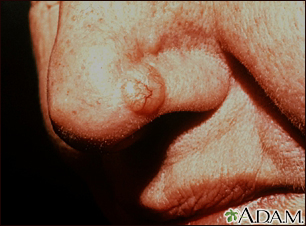
Skin cancer, basal cell carcinoma - nose - illustration
The typical basal cell skin cancer appears as a small, pearly, dome-shaped nodule with small visible blood vessels (telangiectasias).
Skin cancer, basal cell carcinoma - nose
illustration
Skin cancer, basal cell carcinoma - pigmented - illustration
This skin cancer appears as a 2 to 3 centimeter skin spot. The tissue has become destroyed (forming an atrophic plaque). There is a brownish color because of increased skin pigment (hyperpigmentation) and a slightly elevated, rolled, pearl-colored margin. This growth is located along the hair line.
Skin cancer, basal cell carcinoma - pigmented
illustration
Skin cancer, basal cell carcinoma - behind ear - illustration
This skin cancer appears as a 1 to 1.5 centimeter flesh-colored nodule with a central depression and a raised, pearly border. Small blood vessels are visible (telangiectatic).
Skin cancer, basal cell carcinoma - behind ear
illustration
Skin cancer, basal cell carcinoma - spreading - illustration
This skin cancer, a basal cell carcinoma, is 5 to 6 centimeters across, red (erythematous), with well defined (demarcated) borders and sprinkled brown pigment along the margins. This cancer is located on the person's back.
Skin cancer, basal cell carcinoma - spreading
illustration
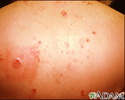
Multiple Basal cell cancer due to x-ray therapy for acne - illustration
Basal cell carcinomas are more prevalent on sun or radiation exposed areas of skin. Here the typical lesion with raised, rolled, pearly borders with ulcerated center is seen on the back of a person previously irradiated for acne.
Multiple Basal cell cancer due to x-ray therapy for acne
illustration
Basal Cell Carcinoma - face - illustration
This flesh-colored, verrucal, pearly, smooth, non-scaling papule is a basal cell carcinoma. It is occurring in a common, sun-exposed area of the face, on the forehead.
Basal Cell Carcinoma - face
illustration
Basal cell carcinoma - close-up - illustration
This basal cell carcinoma appears as a multicolored flat lesion, with a periphery that has ulcerated and bled. Telangiectasia are present.
Basal cell carcinoma - close-up
illustration
Basal cell cancer - illustration
Basal cell cancer is a malignant skin tumor involving cancerous changes of basal skin cells. Basal cell skin cancers usually occur on areas of skin that are regularly exposed to sunlight or other ultraviolet radiation. Once a suspicious lesion is found, a biopsy is needed to prove the diagnosis of basal cell carcinoma. Treatment varies depending on the size, depth, and location of the cancer. Early treatment by a dermatologist may result in a cure rate of more than 95%, but regular examination by a health care provider is required to watch for new sites of basal cell cancer.
Basal cell cancer
illustration


Skin cancer, basal cell carcinoma - nose - illustration
The typical basal cell skin cancer appears as a small, pearly, dome-shaped nodule with small visible blood vessels (telangiectasias).
Skin cancer, basal cell carcinoma - nose
illustration
Skin cancer, basal cell carcinoma - pigmented - illustration
This skin cancer appears as a 2 to 3 centimeter skin spot. The tissue has become destroyed (forming an atrophic plaque). There is a brownish color because of increased skin pigment (hyperpigmentation) and a slightly elevated, rolled, pearl-colored margin. This growth is located along the hair line.
Skin cancer, basal cell carcinoma - pigmented
illustration
Skin cancer, basal cell carcinoma - behind ear - illustration
This skin cancer appears as a 1 to 1.5 centimeter flesh-colored nodule with a central depression and a raised, pearly border. Small blood vessels are visible (telangiectatic).
Skin cancer, basal cell carcinoma - behind ear
illustration
Skin cancer, basal cell carcinoma - spreading - illustration
This skin cancer, a basal cell carcinoma, is 5 to 6 centimeters across, red (erythematous), with well defined (demarcated) borders and sprinkled brown pigment along the margins. This cancer is located on the person's back.
Skin cancer, basal cell carcinoma - spreading
illustration
Multiple Basal cell cancer due to x-ray therapy for acne - illustration
Basal cell carcinomas are more prevalent on sun or radiation exposed areas of skin. Here the typical lesion with raised, rolled, pearly borders with ulcerated center is seen on the back of a person previously irradiated for acne.
Multiple Basal cell cancer due to x-ray therapy for acne
illustration
Basal Cell Carcinoma - face - illustration
This flesh-colored, verrucal, pearly, smooth, non-scaling papule is a basal cell carcinoma. It is occurring in a common, sun-exposed area of the face, on the forehead.
Basal Cell Carcinoma - face
illustration
Basal cell carcinoma - close-up - illustration
This basal cell carcinoma appears as a multicolored flat lesion, with a periphery that has ulcerated and bled. Telangiectasia are present.
Basal cell carcinoma - close-up
illustration
Basal cell cancer - illustration
Basal cell cancer is a malignant skin tumor involving cancerous changes of basal skin cells. Basal cell skin cancers usually occur on areas of skin that are regularly exposed to sunlight or other ultraviolet radiation. Once a suspicious lesion is found, a biopsy is needed to prove the diagnosis of basal cell carcinoma. Treatment varies depending on the size, depth, and location of the cancer. Early treatment by a dermatologist may result in a cure rate of more than 95%, but regular examination by a health care provider is required to watch for new sites of basal cell cancer.
Basal cell cancer
illustration
Review Date: 8/2/2025
Reviewed By: Ramin Fathi, MD, FAAD, Director, Phoenix Surgical Dermatology Group, Phoenix, AZ. Also reviewed by David C. Dugdale, MD, Medical Director, Brenda Conaway, Editorial Director, and the A.D.A.M. Editorial team.
 All rights reserved.