


Retroperitoneal fibrosis
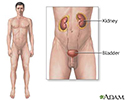
Idiopathic retroperitoneal fibrosis; Ormond's diseaseRetroperitoneal fibrosis is a rare disorder that blocks the tubes (ureters) that carry urine from the kidneys to the bladder.
Causes
Retroperitoneal fibrosis occurs when extra fibrous tissue forms in the area behind the stomach and intestines. The tissue forms a mass (or masses) or tough fibrotic tissue. It can block the tubes that carry urine from the kidney to the bladder.
The cause of this problem is mostly unknown. It is most common in people aged 40 to 60. Men are twice as likely to develop the condition as women.
Symptoms
Early symptoms:
- Dull pain in the abdomen or flank that increases with time
- Pain and change of color in the legs (due to decreased blood flow)
- Swelling of one leg
Later symptoms:
- Decreased urine output
Decreased urine output
Decreased urine output means that you produce less urine than normal. Most adults make at least 500 milliliters of urine in 24 hours (a little over ...
 ImageRead Article Now Book Mark Article
 - No urine output (anuria)
- Nausea, vomiting, changes in mental status caused by kidney failure and build-up of toxic chemicals in the blood
Nausea
Nausea is feeling an urge to vomit. It is often called "being sick to your stomach. "Vomiting or throwing-up forces the contents of the stomach up t...
 ImageRead Article Now Book Mark Article
 - Severe abdominal pain with blood in the stool (due to death of intestinal tissue)
Exams and Tests
Abdominal CT scan is the best way to find a retroperitoneal mass.
Abdominal CT scan
An abdominal CT scan is an imaging test that uses x-rays to create cross-sectional pictures of the belly area. CT stands for computed tomography....

Other tests that can help diagnose this condition include:
- BUN and creatinine blood tests
BUN
BUN stands for blood urea nitrogen. Urea nitrogen is what forms when protein breaks down. A test can be done to measure the amount of urea nitrogen ...
 ImageRead Article Now Book Mark Article
Creatinine blood tests
The creatinine blood test measures the level of creatinine in the blood. This test is done to see how well your kidneys are working. Creatinine in t...
 ImageRead Article Now Book Mark Article
 - Intravenous pyelogram (IVP), not as commonly used
Intravenous pyelogram
An intravenous pyelogram (IVP) is a special x-ray exam of the kidneys, bladder, and ureters (the tubes that carry urine from the kidneys to the bladd...
 ImageRead Article Now Book Mark Article
 - Kidney ultrasound
Kidney ultrasound
Abdominal ultrasound is a type of imaging test. It is used to look at organs in the abdomen, including the liver, gallbladder, pancreas, and kidneys...
 ImageRead Article Now Book Mark Article
 - MRI of the abdomen
MRI of the abdomen
An abdominal magnetic resonance imaging scan is an imaging test that uses powerful magnets and radio waves. The waves create pictures of the inside ...
ImageRead Article Now Book Mark Article - CT scan of the abdomen and retroperitoneum
CT scan of the abdomen
An abdominal CT scan is an imaging test that uses x-rays to create cross-sectional pictures of the belly area. CT stands for computed tomography....
ImageRead Article Now Book Mark Article
A biopsy of the mass may also be done to check for cancer.
Treatment
Corticosteroids are tried first. Some health care providers also prescribe a medicine called tamoxifen.
If corticosteroid treatment does not work, a biopsy should be done to confirm the diagnosis. Other medicines to suppress the immune system can be prescribed.
When medicine does not work, surgery and stents (draining tubes) are needed.
Stents
A stent is a tiny tube placed into a hollow structure in your body. This structure can be an artery, a vein, or another structure, such as the tube ...

Outlook (Prognosis)
The outlook will depend on the extent of the problem and the amount of damage to the kidneys.
The kidney damage may be temporary or permanent.
Kidney damage
Injury to the kidney and ureter is damage to the organs of the upper urinary tract.
Possible Complications
The disorder may lead to:
- Ongoing blockage of the tubes leading from the kidney on one or both sides
- Chronic kidney disease or failure
Chronic kidney disease or failure
Chronic kidney disease (CKD) usually causes the slow loss of kidney function over time. The main job of the kidneys is to remove wastes and excess w...
ImageRead Article Now Book Mark Article
When to Contact a Medical Professional
Contact your provider if you have lower abdomen or flank pain and less output of urine.
Prevention
Try to avoid long-term use of medicines that contain methysergide, ergot alkaloids, or dopamine agonists. These medicines have been shown to rarely cause retroperitoneal fibrosis.
References
Comperat E, Bonsib SM, Cheng L. Renal pelvis and ureter. In: Cheng L, MacLennan GT, Bostwick DG, eds. Urologic Surgical Pathology. 4th ed. Philadelphia, PA: Elsevier; 2020:chap 3.
Nakada SY, Best SL. Management of upper urinary tract obstruction. In: Partin AW, Dmochowski RR, Kavoussi LR, Peters CA, eds. Campbell-Walsh-Wein Urology. 12th ed. Philadelphia, PA: Elsevier; 2021:chap 89.
Privratsky AM, Barreto JC, Tumage RH. Abdominal wall, umbilicus, peritoneum, mesenteries, omentum, and retroperitoneum. In: Townsend CM Jr, Beauchamp RD, Evers BM, Mattox KL, eds. Sabiston Textbook of Surgery. 21st ed. St Louis, MO: Elsevier; 2022:chap 44.
Shanmugam VK. Vasculitis and other uncommon arteriopathies. In: Sidawy AN, Perler BA, eds. Rutherford's Vascular Surgery and Endovascular Therapy. 10th ed. Philadelphia, PA: Elsevier; 2023:chap 138.
Varga J. Systemic sclerosis (scleroderma). In: Goldman L, Cooney KA, eds. Goldman-Cecil Medicine. 27th ed. Philadelphia, PA: Elsevier; 2024:chap 246.
Review Date: 4/1/2025
Reviewed By: Kelly L. Stratton, MD, FACS, Associate Professor, Department of Urology, University of Oklahoma Health Sciences Center, Oklahoma City, OK. Also reviewed by David C. Dugdale, MD, Medical Director, Brenda Conaway, Editorial Director, and the A.D.A.M. Editorial team.
 All rights reserved.