


Pericarditis - after heart attack
Dressler syndrome; Post-MI pericarditis; Post-cardiac injury syndrome; Postcardiotomy pericarditisPericarditis is inflammation and swelling of the covering of the heart (pericardium). It can occur in the days or weeks following a heart attack.
Heart attack
Most heart attacks are caused by a blood clot that blocks one of the coronary arteries. The coronary arteries bring blood and oxygen to the heart. ...


Causes
Two types of pericarditis can occur after a heart attack.
Pericarditis
Pericarditis is a condition in which the sac-like covering around the heart (pericardium) becomes inflamed.

Early pericarditis: This form most often occurs within 1 to 3 days after a heart attack. Inflammation and swelling develop as the body tries to clean up the damaged heart tissue.
Late pericarditis: This is also called Dressler syndrome. It is also called post-cardiac injury syndrome or postcardiotomy pericarditis. It most often develops several weeks or months after a heart attack, heart surgery, or other trauma to the heart. It can also happen about a week after a heart injury. Dressler syndrome is thought to occur when the immune system attacks healthy heart tissue by mistake.
Things that put you at higher risk for pericarditis include:
- A previous heart attack
- Open heart surgery
Open heart surgery
Heart surgery is any surgery done on the heart muscle, valves, arteries, or the aorta and other large arteries connected to the heart. The term "ope...
Read Article Now Book Mark Article - Chest trauma
- A heart attack that has affected the full thickness of your heart muscle
Symptoms
Symptoms include:
- Anxiety
Anxiety
Stress is a feeling of emotional or physical tension. It can come from any event or thought that makes you feel frustrated, angry, or nervous. Stres...
 ImageRead Article Now Book Mark Article
 - Chest pain from the swollen pericardium rubbing on the heart. The pain may be sharp, tight or crushing and may move to the neck, shoulder, or abdomen. The pain may also be worse when you breathe and go away when you lean forward, stand, or sit up.
Chest pain
Chest pain is discomfort or pain that you feel anywhere along the front of your body between your neck and upper abdomen.
 ImageRead Article Now Book Mark Article
 - Trouble breathing
Trouble breathing
Breathing difficulty may involve:Difficult breathing Uncomfortable breathingFeeling like you are not getting enough air
 ImageRead Article Now Book Mark Article
 - Dry cough
Cough
Coughing is an important way to keep your throat and airways clear. But too much coughing may mean you have a disease or disorder. Some coughs are d...
ImageRead Article Now Book Mark Article - Fast heart rate (tachycardia)
Fast heart rate
A bounding pulse is a strong throbbing felt over one of the arteries in the body. It is due to a forceful heartbeat.
 ImageRead Article Now Book Mark Article
 - Fatigue
Fatigue
Fatigue is a feeling of weariness, tiredness, or lack of energy.
 ImageRead Article Now Book Mark Article
 - Fever (common with the second type of pericarditis)
Fever
Fever is the temporary increase in the body's temperature in response to a disease or illness. A child has a fever when the temperature is at or abov...
 ImageRead Article Now Book Mark Article
 - Malaise (general ill feeling)
Malaise
Malaise is a general feeling of discomfort, illness, or lack of well-being.
 ImageRead Article Now Book Mark Article
 - Splinting of ribs (bending over or holding the chest) with deep breathing
Exams and Tests
The health care provider will listen to your heart and lungs with a stethoscope. There may be a rubbing sound (called a pericardial friction rub, not to be confused with a heart murmur). Heart sounds in general may be weak or sound far away.
Heart sounds
A heart murmur is a blowing, whooshing, or rasping sound heard during a heartbeat. The sound is caused by turbulent (rough) blood flow through the h...

A buildup of fluid in the covering of the heart or space around the lungs (pericardial effusion) is not common after a heart attack. But, it often does occur in some people with Dressler syndrome.
Tests may include:
- Cardiac injury markers (CK-MB and troponin may help tell pericarditis from a heart attack)
Troponin
A troponin test measures the levels of troponin T or troponin I proteins in the blood. These proteins are released when the heart muscle has been da...
 ImageRead Article Now Book Mark Article
Pericarditis
Pericarditis is a condition in which the sac-like covering around the heart (pericardium) becomes inflamed.
ImageRead Article Now Book Mark Article - Chest CT scan
Chest CT scan
A chest CT (computed tomography) scan is an imaging method that uses x-rays to create cross-sectional pictures of the chest and upper abdomen....
 ImageRead Article Now Book Mark Article
 - Chest MRI
Chest MRI
A chest MRI (magnetic resonance imaging) scan is an imaging test that uses powerful magnetic fields and radio waves to create pictures of the chest (...
 ImageRead Article Now Book Mark Article
 - Chest x-ray
Chest x-ray
A chest x-ray is an x-ray of the chest, lungs, heart, large arteries, ribs, and diaphragm.
 ImageRead Article Now Book Mark Article
 - Complete blood count (CBC)
Complete blood count
A complete blood count (CBC) test measures the following:The number of white blood cells (WBC count)The number of red blood cells (RBC count)The numb...
 ImageRead Article Now Book Mark Article
 - Electrocardiogram (ECG)
Electrocardiogram
An electrocardiogram (ECG) is a test that records the electrical activity of the heart.
 ImageRead Article Now Book Mark Article
 - Echocardiogram
Echocardiogram
An echocardiogram is a test that uses sound waves to create pictures of the heart. The picture and information it produces is more detailed than a s...
 ImageRead Article Now Book Mark Article
 - ESR (sedimentation rate) or C-reactive protein (measures of inflammation)
ESR
ESR stands for erythrocyte sedimentation rate. It is commonly called a "sed rate. "It is a test that indirectly measures the level of certain protei...
ImageRead Article Now Book Mark ArticleC-reactive protein
C-reactive protein (CRP) is produced by the liver. The level of CRP rises when there is inflammation in the body. It is one of a group of proteins,...
ImageRead Article Now Book Mark Article
Treatment
The goal of treatment is to make the heart work better and reduce pain and other symptoms.
Aspirin may be used to treat inflammation of the pericardium. A drug called colchicine is often used as well.
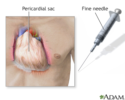
In some cases, excess fluid surrounding the heart (pericardial effusion) may need to be removed. This is done with a procedure called pericardiocentesis. If complications develop, part of the pericardium may sometimes need to be removed with surgery (pericardiectomy).
Pericardiocentesis
Pericardiocentesis is a procedure that uses a needle to remove fluid from the pericardial sac. This is the tissue that surrounds the heart.

Outlook (Prognosis)
The condition may recur in some cases.
Possible Complications
Possible complications of pericarditis are:
- Cardiac tamponade
Cardiac tamponade
Cardiac tamponade is pressure on the heart that occurs when blood or fluid builds up in the space between the heart muscle and the outer covering sac...
ImageRead Article Now Book Mark Article - Congestive heart failure
Congestive heart failure
Heart failure is a condition in which the heart is no longer able to pump oxygen-rich blood to the rest of the body efficiently. This causes symptom...
ImageRead Article Now Book Mark Article - Constrictive pericarditis
Constrictive pericarditis
Constrictive pericarditis is a process in which the sac-like covering of the heart (the pericardium) becomes thickened and scarred. Related conditio...
ImageRead Article Now Book Mark Article
When to Contact a Medical Professional
Contact your provider if:
- You develop symptoms of pericarditis after a heart attack
- You have been diagnosed with pericarditis and symptoms continue or come back despite treatment
References
Hoit BD, Oh JK. Pericardial diseases. In: Goldman L, Cooney KA, eds. Goldman-Cecil Medicine. 27th ed. Philadelphia, PA: Elsevier; 2024:chap 62.
Jouriles NJ. Pericardial and myocardial disease. In: Libby P, Bonow RO, Mann DL, Tomaselli, GF, Bhatt DL, Solomon SD, eds. Braunwald's Heart Disease: A Textbook of Cardiovascular Medicine. 12th ed. Philadelphia, PA: Elsevier; 2022:chap 68.
Lewinter MM, Cremer PC, Klein AL. Pericardial diseases. In: Libby P, Bonow RO, Mann DL, Tomaselli, GF, Bhatt DL, Solomon SD, eds. Braunwald's Heart Disease: A Textbook of Cardiovascular Medicine. 12th ed. Philadelphia, PA: Elsevier; 2022:chap 86.
Acute MI - illustration
A heart attack or acute myocardial infarction (MI) occurs when one of the arteries that supplies the heart muscle becomes blocked. Blockage may be caused by spasm of the artery or by atherosclerosis with acute clot formation. The blockage results in damaged tissue and a permanent loss of contraction of this portion of the heart muscle.
Acute MI
illustration
Pericardium - illustration
The pericardium is a thin double-layered sac which encloses the heart. Fluid is contained within the layers and lubricates the constantly rubbing surfaces.
Pericardium
illustration
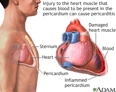
Pericarditis post-IM - illustration
Post-MI pericarditis is inflammation of the pericardium (the sac-like covering of the heart). Any previous injury to the heart muscle can cause pericarditis. Pain occurs when the inflamed pericardium rubs on the heart.
Pericarditis post-IM
illustration
Pericardium - illustration
The pericardial sac surrounds and protects the heart within the chest cavity.
Pericardium
illustration


Acute MI - illustration
A heart attack or acute myocardial infarction (MI) occurs when one of the arteries that supplies the heart muscle becomes blocked. Blockage may be caused by spasm of the artery or by atherosclerosis with acute clot formation. The blockage results in damaged tissue and a permanent loss of contraction of this portion of the heart muscle.
Acute MI
illustration
Pericardium - illustration
The pericardium is a thin double-layered sac which encloses the heart. Fluid is contained within the layers and lubricates the constantly rubbing surfaces.
Pericardium
illustration
Pericarditis post-IM - illustration
Post-MI pericarditis is inflammation of the pericardium (the sac-like covering of the heart). Any previous injury to the heart muscle can cause pericarditis. Pain occurs when the inflamed pericardium rubs on the heart.
Pericarditis post-IM
illustration
Pericardium - illustration
The pericardial sac surrounds and protects the heart within the chest cavity.
Pericardium
illustration
Review Date: 7/14/2024
Reviewed By: Michael A. Chen, MD, PhD, Associate Professor of Medicine, Division of Cardiology, Harborview Medical Center, University of Washington Medical School, Seattle, WA. Also reviewed by David C. Dugdale, MD, Medical Director, Brenda Conaway, Editorial Director, and the A.D.A.M. Editorial team.
 All rights reserved.