


Lung cancer - small cell
Cancer - lung - small cell; Small cell lung cancer; SCLCSmall cell lung cancer (SCLC) is a fast-growing type of lung cancer. It tends to spread more quickly than non-small cell lung cancer.
Lung cancer
Lung cancer is cancer that starts in the lungs. The lungs are located in the chest. When you breathe, air goes through your nose, down your windpipe...

Non-small cell lung cancer
Non-small cell lung cancer is the most common type of lung cancer. It usually grows and spreads more slowly than small cell lung cancer. There are t...
There are two types of SCLC:
- Small cell carcinoma (oat cell cancer)
- Combined small cell carcinoma and non-small cell lung cancer
Most SCLCs are of the oat cell type.
Causes
About 15% of all lung cancer cases are SCLC. Small cell lung cancer is slightly more common in men than women.
Almost all cases of SCLC are due to cigarette smoking. SCLC is very rare in people who have never smoked.
SCLC is the most aggressive form of lung cancer. It usually starts in the breathing tubes (bronchi) in the center of the chest. Although the cancer cells are small, they grow very quickly and create large tumors. These tumors often spread rapidly (metastasize) to other parts of the body, including the brain, liver, and bone.
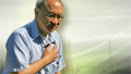
Symptoms
Symptoms of SCLC include:
- Bloody sputum (phlegm)
Bloody sputum
Coughing up blood is the spitting up of blood or bloody mucus from the lungs and throat (respiratory tract). Hemoptysis is the medical term for cough...
 ImageRead Article Now Book Mark Article
 - Chest pain
Chest pain
Chest pain is discomfort or pain that you feel anywhere along the front of your body between your neck and upper abdomen.
 ImageRead Article Now Book Mark Article
 - Cough
- Loss of appetite
- Shortness of breath
Shortness of breath
Breathing difficulty may involve:Difficult breathing Uncomfortable breathingFeeling like you are not getting enough air
ImageRead Article Now Book Mark Article - Weight loss
Weight loss
Unexplained weight loss is a decrease in body weight, when you did not try to lose the weight on your own. Many people gain and lose weight. Uninten...
Read Article Now Book Mark Article - Wheezing
Wheezing
Wheezing is a high-pitched whistling sound during breathing. It occurs when air moves through narrowed breathing tubes in the lungs.
ImageRead Article Now Book Mark Article
Other symptoms that may occur with this disease, especially in the late stages, include:
- Facial swelling
Facial swelling
Facial swelling is the buildup of fluid in the tissues of the face. Swelling may also affect the neck and upper arms.
 ImageRead Article Now Book Mark Article
 - Fever
- Hoarseness or changing voice
Hoarseness or changing voice
Hoarseness refers to difficulty making sounds when trying to speak. Vocal sounds may be weak, breathy, scratchy, or husky, and the pitch or quality ...
 ImageRead Article Now Book Mark Article
 - Swallowing difficulty
Swallowing difficulty
Difficulty with swallowing is the feeling that food or liquid is stuck in the throat or at any point before the food enters the stomach. This proble...
 ImageRead Article Now Book Mark Article
 - Weakness

Exams and Tests
Your health care provider will perform a physical exam and ask about your medical history. You will be asked whether you smoke, and if so, how much and for how long.
When listening to your chest with a stethoscope, your provider may hear fluid around the lungs or areas where the lung has partially collapsed. Each of these findings may suggest cancer.
Collapsed
A collapsed lung occurs when air escapes from the lung. The air then can fill the space outside of the lung between the lung and chest wall. This b...
SCLC has usually spread to other parts of your body by the time it is diagnosed.
Tests that may be performed include:
- Bone scan
Bone scan
A bone scan is an imaging test used to diagnose bone diseases and find out how severe they are.
 ImageRead Article Now Book Mark Article
 - Chest x-ray
Chest x-ray
A chest x-ray is an x-ray of the chest, lungs, heart, large arteries, ribs, and diaphragm.
 ImageRead Article Now Book Mark Article
 - Complete blood count (CBC)
CBC
A complete blood count (CBC) test measures the following:The number of white blood cells (WBC count)The number of red blood cells (RBC count)The numb...
 ImageRead Article Now Book Mark Article
 - CT scan
CT scan
A chest CT (computed tomography) scan is an imaging method that uses x-rays to create cross-sectional pictures of the chest and upper abdomen....
 ImageRead Article Now Book Mark Article
 - Liver function tests
Liver function tests
Liver function tests are common tests that are used to see how well the liver is working. Tests include:AlbuminAlpha-1 antitrypsinAlkaline phosphata...
 ImageRead Article Now Book Mark Article
 - MRI scan
MRI scan
A chest MRI (magnetic resonance imaging) scan is an imaging test that uses powerful magnetic fields and radio waves to create pictures of the chest (...
 ImageRead Article Now Book Mark Article
 - Positron emission tomography (PET) scan
PET
A lung positron emission tomography (PET) scan is an imaging test. It uses a radioactive substance (called a tracer) to look for disease in the lung...
ImageRead Article Now Book Mark Article - Sputum cytology test (to look for cancer cells), seldom done today
Sputum cytology test
Routine sputum culture is a laboratory test that looks for germs that cause infection. Sputum is the material that comes up from air passages when y...
 ImageRead Article Now Book Mark Article
 - Thoracentesis (removal of fluid from the chest cavity around the lungs)
Thoracentesis
Thoracentesis is a procedure to remove fluid from the space between the lining of the outside of the lungs (pleura) and the wall of the chest....
Read Article Now Book Mark Article
In most cases, a piece of tissue is removed from your lungs or other areas to be examined under a microscope. This is called a biopsy. There are several ways to do a biopsy:
- Bronchoscopy combined with biopsy
Bronchoscopy
Bronchoscopy is a test to view the airways and diagnose lung disease. It may also be used during the treatment of some lung conditions.
 ImageRead Article Now Book Mark Article
Biopsy
A biopsy is the removal of a small piece of tissue for lab examination.
 ImageRead Article Now Book Mark Article
 - CT scan-directed needle biopsy
CT scan-directed needle biopsy
A lung needle biopsy is a method to remove a piece of lung tissue for examination. If it is done through the wall of your chest, it is called a tran...
 ImageRead Article Now Book Mark Article
 - Endoscopic esophageal or bronchial ultrasound with biopsy
- Mediastinoscopy with biopsy
Mediastinoscopy with biopsy
Mediastinoscopy with biopsy is a procedure in which a lighted instrument (mediastinoscope) is inserted in the space in the chest between the lungs (m...
 ImageRead Article Now Book Mark Article
 - Open lung biopsy
Open lung biopsy
An open lung biopsy is surgery to remove a small piece of tissue from the lung. The sample is then examined for cancer, infection, or lung disease....
ImageRead Article Now Book Mark Article - Pleural biopsy
Pleural biopsy
Pleural biopsy is a procedure to remove a sample of the pleura. This is the thin tissue that lines the chest cavity and surrounds the lungs. The bi...
 ImageRead Article Now Book Mark Article
 - Video-assisted thoracoscopy
Video-assisted thoracoscopy
Lung surgery is surgery done to repair or remove lung tissue. There are many common lung surgeries, including:Biopsy of an unknown growth in or arou...
 ImageRead Article Now Book Mark Article
Usually, if a biopsy shows cancer, more imaging tests are done to find out the stage of the cancer. Stage means how far it has spread. SCLC is classified as either:
- Limited -- Cancer is only in the chest and can be treated with radiation therapy.
- Extensive -- Cancer has spread outside the area that can be covered by radiation therapy.
Treatment
Because SCLC spreads quickly throughout the body, treatment will include cancer-killing medicines (chemotherapy), which are usually given through a vein (by IV).
Chemotherapy
The term chemotherapy is used to describe cancer-killing drugs. Chemotherapy may be used to:Cure the cancerShrink the cancerPrevent the cancer from ...

Treatment with chemotherapy and immunotherapy, and possibly radiation, may be done for people with SCLC that has spread throughout the body (extensive). In this case, the treatment only helps relieve symptoms and prolongs life, but does not cure the disease.
Radiation
Radiation therapy uses high-powered radiation (such as x-rays or gamma rays), particles, or radioactive seeds to kill cancer cells.

Radiation therapy can be used with chemotherapy if the disease is confined to one area within the chest (limited).
Radiation therapy uses powerful x-rays or other forms of radiation to kill cancer cells.
Radiation may be used to:
- Treat the cancer, along with chemotherapy, if surgery is not possible.
- Help relieve symptoms caused by the cancer, such as breathing problems and swelling.
- Help relieve cancer pain when the cancer has spread to the bones.
Often, SCLC may have already spread to the brain. This can occur even when there are no symptoms or other signs of cancer in the brain. As a result, some people with smaller cancers, or who had a good response in their first round of chemotherapy, may receive radiation therapy to the brain. This therapy, called prophylactic cranial irradiation, is done to prevent growth of the cancer in the brain.
Surgery helps very few people with SCLC because the disease has often spread by the time it is diagnosed. Surgery may be done when there is only one tumor that has not spread. If surgery is done, chemotherapy or radiation therapy is still needed.
Support Groups
You can ease the stress of illness by joining a cancer support group. Sharing with others who have common experiences and problems can help you not feel alone.
Cancer support group
The following organizations are good resources for information on cancer:American Cancer Society. Support and online communities. www. cancer. org/...
Outlook (Prognosis)
How well you do depends on how much the lung cancer has spread. SCLC is very deadly. Not many people with this type of cancer are still alive 5 years after diagnosis.
Treatment can often prolong life to more than 12 months, even when the cancer has spread.
In rare cases, if SCLC is diagnosed early, treatment may result in a long-term cure.
When to Contact a Medical Professional
Contact your provider if you have symptoms of lung cancer, particularly if you smoke.
Prevention
If you smoke, now is the time to quit. If you are having trouble quitting, talk with your provider. There are many methods to help you quit, from support groups to prescription medicines. Also try to avoid secondhand smoke.
Trouble quitting
There are many ways to quit smoking. There are also resources to help you. Family members, friends, and co-workers may be supportive. But to be su...

Support groups
It is hard to quit smoking if you are acting alone. Smokers usually have a much better chance of quitting with a support program. Stop smoking prog...

Prescription medicines
Nicotine replacement therapy is a treatment to help people stop smoking. It uses products that supply low doses of nicotine. These products do not ...
If you smoke or used to smoke, talk with your provider about getting screened for lung cancer. To get screened, you need to have a CT scan of the chest.
References
Araujo LH, Horn L, Merritt RE, Shilo K, Xu-Welliver M, Carbone DP. Cancer of the lung: non-small cell lung cancer and small cell lung cancer. In: Niederhuber JE, Armitage JO, Kastan MB, Doroshow JH, Tepper JE, eds. Abeloff's Clinical Oncology. 6th ed. Philadelphia, PA: Elsevier; 2020:chap 69.
National Comprehensive Cancer Network website. NCCN clinical practice guidelines in oncology: Small cell lung cancer. Version 4. 2025. www.nccn.org/professionals/physician_gls/pdf/sclc.pdf. Updated January 13, 2025. Accessed July 18, 2025.
National Cancer Institute website. Small cell lung cancer treatment (PDQ) - health professional version. www.cancer.gov/types/lung/hp/small-cell-lung-treatment-pdq. Updated May 14, 2025. Accessed July 18, 2025.
Rivera MP, Mody GN, Weiner AA. Lung cancer: treatment. In: Broaddus VC, Ernst JD, Talmadge EK, et al, eds. Murray and Nadel's Textbook of Respiratory Medicine. 7th ed. Philadelphia, PA: Elsevier; 2022:chap 77.
Bronchoscopy - illustration
Bronchoscopy is a surgical technique for viewing the interior of the airways. Using sophisticated flexible fiber optic instruments, surgeons are able to explore the trachea, main stem bronchi, and some of the small bronchi. In children, this procedure may be used to remove foreign objects that have been inhaled. In adults, the procedure is most often used to take samples of (biopsy) suspicious lesions and for culturing specific areas in the lung.
Bronchoscopy
illustration
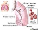
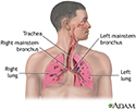
Lungs - illustration
The major features of the lungs include the bronchi, the bronchioles and the alveoli. The alveoli are the microscopic blood vessel-lined sacks in which oxygen and carbon dioxide gas are exchanged.
Lungs
illustration
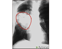
Lung cancer - lateral chest x-ray - illustration
A lateral view of a chest x-ray in a patient with central cancer of the lung.
Lung cancer - lateral chest x-ray
illustration
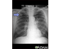
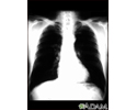
Lung cancer - frontal chest X-ray - illustration
A chest x-ray in a patient with central cancer of the right lung. Notice the white mass in the middle portion of the right lung (seen on the left side of the picture).
Lung cancer - frontal chest X-ray
illustration
Adenocarcinoma - chest x-ray - illustration
This chest x-ray shows adenocarcinoma of the lung. There is a rounded light spot in the right upper lung (left side of the picture) at the level of the second rib. The light spot has irregular and poorly defined borders and is not uniform in density. Diseases that may cause this type of x-ray result would be tuberculous or fungal granuloma, and malignant or benign tumors.
Adenocarcinoma - chest x-ray
illustration
Bronchial cancer - CT scan - illustration
This chest CT scan shows a cross-section of a person with bronchial cancer. The two dark areas are the lungs. The light areas within the lungs represent the cancer.
Bronchial cancer - CT scan
illustration
Bronchial cancer - chest X-ray - illustration
This is a chest x-ray of a person with bronchial cancer. This is a front view. The lungs are the two dark areas. The heart and other structures are white areas visible in the middle of the chest. The light areas that appear as subtle branches extending from the center into the lungs are cancerous.
Bronchial cancer - chest X-ray
illustration
Lung with squamous cell cancer - CT scan - illustration
This CT scan shows a cross section of the lungs of a person with lung cancer. The two dark areas in the middle of the screen are the lungs. The light areas in the right lung (on the left of the screen) represent the cancer.
Lung with squamous cell cancer - CT scan
illustration
Lung cancer - chemotherapy treatment - illustration
Treatment for lung cancer depends on the type of cancer and the stage of the disease. Chemotherapy is a form of treatment for lung cancer which may cure, shrink or keep the cancer from spreading.
Lung cancer - chemotherapy treatment
illustration
Adenocarcinoma - illustration
Adenocarcinoma usually develops on the outer boundaries of the lungs and is more commonly found in women than in men.
Adenocarcinoma
illustration
Non-small cell carcinoma - illustration
Non-small cell carcinomas are the most common lung cancers.
Non-small cell carcinoma
illustration
Small cell carcinoma - illustration
Small cell carcinoma, also called oat cell carcinoma, can create its own hormones, which alter body chemistry.
Small cell carcinoma
illustration
Squamous cell carcinoma - illustration
Squamous cell carcinoma commonly starts in the bronchi and may not spread as rapidly as other lung cancers.
Squamous cell carcinoma
illustration
Secondhand smoke and lung cancer - illustration
Secondhand smoke has been classified as a known cause of lung cancer in humans (Group A carcinogen).
Secondhand smoke and lung cancer
illustration
Normal lungs and alveoli - illustration
The lungs are located in the chest cavity and are responsible for respiration. The alveoli are small sir sacs where oxygen is exchanged in the lungs.
Normal lungs and alveoli
illustration
Respiratory system - illustration
Air is breathed in through the nasal passageways, travels through the trachea and bronchi to the lungs.
Respiratory system
illustration
Smoking hazards - illustration
Smoking cigarettes puts a person at risk for emphysema, lung cancer and other health problems. Second-hand smoke (smoke from a nearby cigarette) may also contribute to these diseases, especially among children.
Smoking hazards
illustration
Bronchoscope - illustration
Lung or bronchial biopsy (tissue samples taken to diagnose or rule out disease) may be done with the use of a flexible bronchoscope, an instrument with a small light and camera which is inserted through the nose or mouth. When a suspicious area is seen, an instrument is channeled through the bronchoscope to take a sample for analysis.
Bronchoscope
illustration


Bronchoscopy - illustration
Bronchoscopy is a surgical technique for viewing the interior of the airways. Using sophisticated flexible fiber optic instruments, surgeons are able to explore the trachea, main stem bronchi, and some of the small bronchi. In children, this procedure may be used to remove foreign objects that have been inhaled. In adults, the procedure is most often used to take samples of (biopsy) suspicious lesions and for culturing specific areas in the lung.
Bronchoscopy
illustration
Lungs - illustration
The major features of the lungs include the bronchi, the bronchioles and the alveoli. The alveoli are the microscopic blood vessel-lined sacks in which oxygen and carbon dioxide gas are exchanged.
Lungs
illustration
Lung cancer - lateral chest x-ray - illustration
A lateral view of a chest x-ray in a patient with central cancer of the lung.
Lung cancer - lateral chest x-ray
illustration
Lung cancer - frontal chest X-ray - illustration
A chest x-ray in a patient with central cancer of the right lung. Notice the white mass in the middle portion of the right lung (seen on the left side of the picture).
Lung cancer - frontal chest X-ray
illustration
Adenocarcinoma - chest x-ray - illustration
This chest x-ray shows adenocarcinoma of the lung. There is a rounded light spot in the right upper lung (left side of the picture) at the level of the second rib. The light spot has irregular and poorly defined borders and is not uniform in density. Diseases that may cause this type of x-ray result would be tuberculous or fungal granuloma, and malignant or benign tumors.
Adenocarcinoma - chest x-ray
illustration
Bronchial cancer - CT scan - illustration
This chest CT scan shows a cross-section of a person with bronchial cancer. The two dark areas are the lungs. The light areas within the lungs represent the cancer.
Bronchial cancer - CT scan
illustration
Bronchial cancer - chest X-ray - illustration
This is a chest x-ray of a person with bronchial cancer. This is a front view. The lungs are the two dark areas. The heart and other structures are white areas visible in the middle of the chest. The light areas that appear as subtle branches extending from the center into the lungs are cancerous.
Bronchial cancer - chest X-ray
illustration
Lung with squamous cell cancer - CT scan - illustration
This CT scan shows a cross section of the lungs of a person with lung cancer. The two dark areas in the middle of the screen are the lungs. The light areas in the right lung (on the left of the screen) represent the cancer.
Lung with squamous cell cancer - CT scan
illustration
Lung cancer - chemotherapy treatment - illustration
Treatment for lung cancer depends on the type of cancer and the stage of the disease. Chemotherapy is a form of treatment for lung cancer which may cure, shrink or keep the cancer from spreading.
Lung cancer - chemotherapy treatment
illustration
Adenocarcinoma - illustration
Adenocarcinoma usually develops on the outer boundaries of the lungs and is more commonly found in women than in men.
Adenocarcinoma
illustration
Non-small cell carcinoma - illustration
Non-small cell carcinomas are the most common lung cancers.
Non-small cell carcinoma
illustration
Small cell carcinoma - illustration
Small cell carcinoma, also called oat cell carcinoma, can create its own hormones, which alter body chemistry.
Small cell carcinoma
illustration
Squamous cell carcinoma - illustration
Squamous cell carcinoma commonly starts in the bronchi and may not spread as rapidly as other lung cancers.
Squamous cell carcinoma
illustration
Secondhand smoke and lung cancer - illustration
Secondhand smoke has been classified as a known cause of lung cancer in humans (Group A carcinogen).
Secondhand smoke and lung cancer
illustration
Normal lungs and alveoli - illustration
The lungs are located in the chest cavity and are responsible for respiration. The alveoli are small sir sacs where oxygen is exchanged in the lungs.
Normal lungs and alveoli
illustration
Respiratory system - illustration
Air is breathed in through the nasal passageways, travels through the trachea and bronchi to the lungs.
Respiratory system
illustration
Smoking hazards - illustration
Smoking cigarettes puts a person at risk for emphysema, lung cancer and other health problems. Second-hand smoke (smoke from a nearby cigarette) may also contribute to these diseases, especially among children.
Smoking hazards
illustration
Bronchoscope - illustration
Lung or bronchial biopsy (tissue samples taken to diagnose or rule out disease) may be done with the use of a flexible bronchoscope, an instrument with a small light and camera which is inserted through the nose or mouth. When a suspicious area is seen, an instrument is channeled through the bronchoscope to take a sample for analysis.
Bronchoscope
illustration
Review Date: 7/9/2025
Reviewed By: Warren Brenner, MD, Oncologist, Lynn Cancer Institute, Boca Raton, FL. Review provided by VeriMed Healthcare Network. Also reviewed by David C. Dugdale, MD, Medical Director, Brenda Conaway, Editorial Director, and the A.D.A.M. Editorial team.
 All rights reserved.