


Lung metastases
Metastases to the lung; Metastatic cancer to the lung; Lung cancer - metastases; Lung metsLung metastases are cancerous tumors that start somewhere else in the body and spread to the lungs.
Causes
Metastatic tumors in the lungs are cancers that developed at other places in the body (or other parts of the lungs). They then spread through the bloodstream or lymphatic system to the lungs. It is different than lung cancer that starts in the lungs.
Nearly any cancer can spread to the lungs. Common cancers include:
- Bladder cancer
Bladder cancer
Bladder cancer is a cancer that starts in the bladder. The bladder is the body part that holds and releases urine. It is in the center of the lower...
 ImageRead Article Now Book Mark Article
 - Breast cancer
Breast cancer
Breast cancer is cancer that starts in the tissues of the breast. There are two main types of breast cancer:Ductal carcinoma starts in the tubes (du...
 ImageRead Article Now Book Mark Article
 - Lung cancer
- Colorectal cancer
Colorectal cancer
Colorectal cancer is cancer that starts in the large intestine (colon) or the rectum (end of the colon). It is also sometimes simply called colon ca...
 ImageRead Article Now Book Mark Article
 - Kidney cancer
- Melanoma
Melanoma
Melanoma is the most dangerous type of skin cancer. It is also the rarest. It is the leading cause of death from skin disease. Other common types o...
 ImageRead Article Now Book Mark Article
 - Ovarian cancer
Ovarian cancer
Ovarian cancer is cancer that starts in the ovaries. The ovaries are the female reproductive organs that produce eggs.
 ImageRead Article Now Book Mark Article
 - Sarcoma
- Thyroid cancer
Thyroid cancer
Thyroid cancer is a cancer that starts in the thyroid gland. The thyroid gland is located in the front of your lower neck.
 ImageRead Article Now Book Mark Article
 - Pancreatic cancer
Pancreatic cancer
Pancreatic cancer is cancer that starts in the pancreas.
 ImageRead Article Now Book Mark Article
 - Testicular cancer
Testicular cancer
Testicular cancer is cancer that starts in the testicles. The testicles are the male reproductive glands located in the scrotum.
 ImageRead Article Now Book Mark Article
Symptoms
Symptoms may include any of the following:
- Bloody sputum
Bloody sputum
Coughing up blood is the spitting up of blood or bloody mucus from the lungs and throat (respiratory tract). Hemoptysis is the medical term for cough...
 ImageRead Article Now Book Mark Article
 - Chest pain
Chest pain
Chest pain is discomfort or pain that you feel anywhere along the front of your body between your neck and upper abdomen.
 ImageRead Article Now Book Mark Article
 - Cough
Cough
Coughing is an important way to keep your throat and airways clear. But too much coughing may mean you have a disease or disorder. Some coughs are d...
 ImageRead Article Now Book Mark Article
 - Shortness of breath
Shortness of breath
Breathing difficulty may involve:Difficult breathing Uncomfortable breathingFeeling like you are not getting enough air
ImageRead Article Now Book Mark Article - Weakness
- Weight loss
Exams and Tests
Your health care provider will examine you and ask about your medical history and symptoms. Tests that may be done include:
- Bronchoscopy to view the airways and take tissue samples, if possible
Bronchoscopy
Bronchoscopy is a test to view the airways and diagnose lung disease. It may also be used during the treatment of some lung conditions.
 ImageRead Article Now Book Mark Article
 - Chest CT scan
Chest CT scan
A chest CT (computed tomography) scan is an imaging method that uses x-rays to create cross-sectional pictures of the chest and upper abdomen....
 ImageRead Article Now Book Mark Article
 - Chest x-ray
Chest x-ray
A chest x-ray is an x-ray of the chest, lungs, heart, large arteries, ribs, and diaphragm.
 ImageRead Article Now Book Mark Article
 - Cytologic studies of pleural fluid or sputum
Cytologic studies of pleural fluid
A cytology exam of pleural fluid is a lab test to detect cancer cells and certain other cells in the fluid from the area that surrounds the lungs. T...
Read Article Now Book Mark Article - Lung needle biopsy
Lung needle biopsy
A lung needle biopsy is a method to remove a piece of lung tissue for examination. If it is done through the wall of your chest, it is called a tran...
 ImageRead Article Now Book Mark Article
 - PET scan
PET scan
A lung positron emission tomography (PET) scan is an imaging test. It uses a radioactive substance (called a tracer) to look for disease in the lung...
ImageRead Article Now Book Mark Article - Surgery to take a sample of tissue from the lungs (surgical lung biopsy)
Surgical lung biopsy
An open lung biopsy is surgery to remove a small piece of tissue from the lung. The sample is then examined for cancer, infection, or lung disease....
ImageRead Article Now Book Mark Article
Treatment
Chemotherapy is often used to treat metastatic cancer to the lung. Surgery to remove the tumors may be done when any of the following occurs:
- The cancer has spread to only limited areas of the lung
- The lung tumors can be completely removed with surgery
However, the main tumor must be curable, and the person must be strong enough to go through the surgery and recovery.
Other treatments include:
- Radiation therapy
Radiation therapy
Radiation therapy uses high-powered radiation (such as x-rays or gamma rays), particles, or radioactive seeds to kill cancer cells.
 ImageRead Article Now Book Mark Article
 - Immunotherapy
- Targeted systemic therapy
- The placement of stents inside the airways
Stents
A stent is a tiny tube placed into a hollow structure in your body. This structure can be an artery, a vein, or another structure, such as the tube ...
 ImageRead Article Now Book Mark Article
 - Laser therapy
Laser therapy
Laser therapy uses a very narrow, focused beam of light to shrink or destroy cancer cells. It can be used to cut out tumors without damaging other t...
Read Article Now Book Mark Article - Using local heat probes to destroy the cancer
- Using very cold temperature to destroy the cancer
Support Groups
You can ease the stress of illness by joining a support group where members share common experiences and problems.
Support group
The following organizations are good resources for information on cancer:American Cancer Society. Support and online communities. www. cancer. org/...
Outlook (Prognosis)
A cure is unlikely in most cases of cancers that have spread to the lungs. But the outlook depends on the main cancer. In some cases, a person can live more than 5 years with metastatic cancer to the lungs.
Possible Complications
Complications of metastatic tumors in the lungs may include:
- Fluid between the lung and chest wall (pleural effusion), which can cause shortness of breath or pain when taking a deep breath
Pleural effusion
A pleural effusion is a buildup of fluid between the layers of tissue that line the lungs and chest cavity.
ImageRead Article Now Book Mark Article - Further spread of the cancer
When to Contact a Medical Professional
Contact your provider if you have a history of cancer and you develop:
- Coughing up blood
- Persistent cough
- Shortness of breath
- Unexplained weight loss
Prevention
Not all cancers can be prevented. However, many can be prevented by:
- Eating healthy foods
- Exercising regularly
- Limiting alcohol consumption
- Not smoking
References
Arenberg DA, Reddy RM. Metastatic malignant tumors. In: Broaddus VC, Ernst JD, King TE, et al, eds. Murray and Nadel's Textbook of Respiratory Medicine. 7th ed. Philadelphia, PA: Elsevier; 2022:chap 79.
Hayman J, Naidoo J, Ettinger DS. Lung metastases. In: Niederhuber JE, Armitage JO, Kastan MB, Doroshow JH, Tepper JE, eds. Abeloff's Clinical Oncology. 6th ed. Philadelphia, PA: Elsevier; 2020:chap 57.
Wald O, Izhar U, Sugarbaker DJ. Lung, chest wall, pleura, and mediastinum. In: Townsend CM Jr, Beauchamp RD, Evers BM, Mattox KL, eds. Sabiston Textbook of Surgery. 21st ed. St Louis, MO: Elsevier; 2022:chap 58.
Bronchoscopy - illustration
Bronchoscopy is a surgical technique for viewing the interior of the airways. Using sophisticated flexible fiber optic instruments, surgeons are able to explore the trachea, main stem bronchi, and some of the small bronchi. In children, this procedure may be used to remove foreign objects that have been inhaled. In adults, the procedure is most often used to take samples of (biopsy) suspicious lesions and for culturing specific areas in the lung.
Bronchoscopy
illustration
Lung cancer - lateral chest x-ray - illustration
A lateral view of a chest x-ray in a patient with central cancer of the lung.
Lung cancer - lateral chest x-ray
illustration
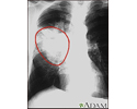
Lung cancer - frontal chest X-ray - illustration
A chest x-ray in a patient with central cancer of the right lung. Notice the white mass in the middle portion of the right lung (seen on the left side of the picture).
Lung cancer - frontal chest X-ray
illustration
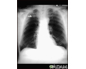
Pulmonary nodule - front view chest x-ray - illustration
This x-ray shows a single lesion (pulmonary nodule) in the upper right lung (seen as a light area on the left side of the picture). The nodule has distinct borders (well-defined) and is uniform in density. Tuberculosis (TB) and other diseases can cause this type of lesion.
Pulmonary nodule - front view chest x-ray
illustration
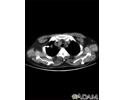
Pulmonary nodule, solitary - CT scan - illustration
This CT scan shows a single lesion (pulmonary nodule) in the right lung. This nodule is seen as the light circle in the upper portion of the dark area on the left side of the picture. A normal lung would look completely black in a CT scan.
Pulmonary nodule, solitary - CT scan
illustration
Lung with squamous cell cancer - CT scan - illustration
This CT scan shows a cross section of the lungs of a person with lung cancer. The two dark areas in the middle of the screen are the lungs. The light areas in the right lung (on the left of the screen) represent the cancer.
Lung with squamous cell cancer - CT scan
illustration
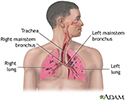
Respiratory system - illustration
Air is breathed in through the nasal passageways, travels through the trachea and bronchi to the lungs.
Respiratory system
illustration


Bronchoscopy - illustration
Bronchoscopy is a surgical technique for viewing the interior of the airways. Using sophisticated flexible fiber optic instruments, surgeons are able to explore the trachea, main stem bronchi, and some of the small bronchi. In children, this procedure may be used to remove foreign objects that have been inhaled. In adults, the procedure is most often used to take samples of (biopsy) suspicious lesions and for culturing specific areas in the lung.
Bronchoscopy
illustration
Lung cancer - lateral chest x-ray - illustration
A lateral view of a chest x-ray in a patient with central cancer of the lung.
Lung cancer - lateral chest x-ray
illustration
Lung cancer - frontal chest X-ray - illustration
A chest x-ray in a patient with central cancer of the right lung. Notice the white mass in the middle portion of the right lung (seen on the left side of the picture).
Lung cancer - frontal chest X-ray
illustration
Pulmonary nodule - front view chest x-ray - illustration
This x-ray shows a single lesion (pulmonary nodule) in the upper right lung (seen as a light area on the left side of the picture). The nodule has distinct borders (well-defined) and is uniform in density. Tuberculosis (TB) and other diseases can cause this type of lesion.
Pulmonary nodule - front view chest x-ray
illustration
Pulmonary nodule, solitary - CT scan - illustration
This CT scan shows a single lesion (pulmonary nodule) in the right lung. This nodule is seen as the light circle in the upper portion of the dark area on the left side of the picture. A normal lung would look completely black in a CT scan.
Pulmonary nodule, solitary - CT scan
illustration
Lung with squamous cell cancer - CT scan - illustration
This CT scan shows a cross section of the lungs of a person with lung cancer. The two dark areas in the middle of the screen are the lungs. The light areas in the right lung (on the left of the screen) represent the cancer.
Lung with squamous cell cancer - CT scan
illustration
Respiratory system - illustration
Air is breathed in through the nasal passageways, travels through the trachea and bronchi to the lungs.
Respiratory system
illustration
Review Date: 6/17/2024
Reviewed By: Todd Gersten, MD, Hematology/Oncology, Florida Cancer Specialists & Research Institute, Wellington, FL. Review provided by VeriMed Healthcare Network. Also reviewed by David C. Dugdale, MD, Medical Director, Brenda Conaway, Editorial Director, and the A.D.A.M. Editorial team.
 All rights reserved.