


Mycoplasma pneumonia
Walking pneumonia; Community-acquired pneumonia - mycoplasma; Community-acquired pneumonia - atypicalPneumonia is inflamed or swollen lung tissue due to infection with a germ.
Mycoplasma pneumonia is caused by the bacteria Mycoplasma pneumoniae (M pneumoniae).
This type of pneumonia is also called atypical pneumonia because the symptoms are different from those of pneumonia due to other common bacteria.
Atypical pneumonia
Pneumonia is inflamed or swollen lung tissue due to infection with a germ. With atypical pneumonia, the infection is caused by different bacteria tha...

Causes
Mycoplasma pneumonia usually affects people younger than 40.
People who live or work in crowded areas such as schools and homeless shelters have a higher chance of getting this condition. But many people who get sick with it have no known risk factors.
Symptoms
Symptoms are often mild and appear over 1 to 3 weeks. They may become more severe in some people.
Common symptoms include any of the following:
- Chest pain
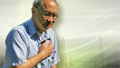
Chest pain
Chest pain is discomfort or pain that you feel anywhere along the front of your body between your neck and upper abdomen.
 ImageRead Article Now Book Mark Article
 - Chills
- Cough, usually dry and not bloody
Cough
Coughing is an important way to keep your throat and airways clear. But too much coughing may mean you have a disease or disorder. Some coughs are d...
ImageRead Article Now Book Mark Article - Excessive sweating
Excessive sweating
Sweating is the release of liquid from the body's sweat glands. This liquid contains salt. This process is also called perspiration. Sweating helps...
 ImageRead Article Now Book Mark Article
 - Fever (may be high)
Fever
Fever is the temporary increase in the body's temperature in response to a disease or illness. A child has a fever when the temperature is at or abov...
 ImageRead Article Now Book Mark Article
 - Headache
Headache
A headache is pain or discomfort in the head, scalp, or neck. Serious causes of headaches are rare. Most people with headaches can feel much better...
 ImageRead Article Now Book Mark Article
 - Sore throat
Less common symptoms include:
- Ear pain
- Eye pain or soreness
Eye pain
Pain in the eye may be described as a burning, throbbing, aching, or stabbing sensation in or around the eye. It may also feel like you have a forei...
 ImageRead Article Now Book Mark Article
 - Muscle aches and joint stiffness
Muscle aches
Muscle aches and pains are common and can involve more than one muscle. Pain due to muscles can also be felt in ligaments, tendons, and fascia. Fas...
 ImageRead Article Now Book Mark Article
 - Neck lump
Neck lump
A neck lump is any lump, bump, or swelling in the neck.
 ImageRead Article Now Book Mark Article
 - Rapid breathing
- Skin lesions or rash
Skin lesions
Rashes involve changes in the color, feeling or texture of your skin.
 ImageRead Article Now Book Mark Article

Exams and Tests
People with suspected pneumonia should have a complete medical evaluation. It may be hard for your health care provider to tell whether you have pneumonia, bronchitis, or another respiratory infection, so you may need a chest x-ray.
Chest x-ray
A chest x-ray is an x-ray of the chest, lungs, heart, large arteries, ribs, and diaphragm.

Depending on how severe your symptoms are, other tests may be done, including:
- Complete blood count (CBC)
Complete blood count
A complete blood count (CBC) test measures the following:The number of white blood cells (WBC count)The number of red blood cells (RBC count)The numb...
 ImageRead Article Now Book Mark Article
 - Blood tests
- Bronchoscopy in which a flexible tube with a lighted camera on the end is passed down to your lungs in selected cases (rarely needed)
Bronchoscopy
Bronchoscopy is a test to view the airways and diagnose lung disease. It may also be used during the treatment of some lung conditions.
 ImageRead Article Now Book Mark Article
 - CT scan of the chest
CT scan of the chest
A chest CT (computed tomography) scan is an imaging method that uses x-rays to create cross-sectional pictures of the chest and upper abdomen....
 ImageRead Article Now Book Mark Article
 - Measuring levels of oxygen and carbon dioxide in the blood (arterial blood gases)
Arterial blood gases
Blood gases are a measurement of how much oxygen and carbon dioxide are in your blood. They also determine the acidity (pH) of your blood.
 ImageRead Article Now Book Mark Article
 - Nose or throat swab to check for bacteria and viruses
- Open lung biopsy (only done in very serious illnesses when the diagnosis cannot be made from other sources, so very rarely needed)
Open lung biopsy
An open lung biopsy is surgery to remove a small piece of tissue from the lung. The sample is then examined for cancer, infection, or lung disease....
ImageRead Article Now Book Mark Article - Sputum tests to check for mycoplasma bacteria
Sputum tests
Routine sputum culture is a laboratory test that looks for germs that cause infection. Sputum is the material that comes up from air passages when y...
 ImageRead Article Now Book Mark Article
In many cases, it is not necessary to make the specific diagnosis before starting treatment.
Treatment
To feel better, you can take these self-care measures at home:
- Control your fever with aspirin, nonsteroidal anti-inflammatory drugs (NSAIDs, such as ibuprofen or naproxen), or acetaminophen. Do not give aspirin to children because it may cause a dangerous illness called Reye syndrome.
Reye syndrome
Reye syndrome is characterized by sudden (acute) brain damage and liver function problems. This condition does not have a known cause. This syndrome...
 ImageRead Article Now Book Mark Article
 - Do not take cough medicines without first contacting your provider. Cough medicines may make it harder for your body to cough up the extra sputum.
- Drink plenty of fluids to help loosen secretions and bring up phlegm.
- Get a lot of rest. Have someone else do household chores.
Antibiotics are used to treat atypical pneumonia:
- You may be able to take antibiotics by mouth at home.
- If your condition is severe, you will likely be admitted to a hospital. There, you will be given antibiotics through a vein (intravenously), as well as oxygen.
Intravenously
Quantitative nephelometry is a lab test to quickly and accurately measure levels of certain proteins called immunoglobulins in the blood. Immunoglob...
 ImageRead Article Now Book Mark Article
 - Antibiotics are usually prescribed for 3 to 5 days, although sometimes they may be used for 2 weeks or more.
- Finish all the antibiotics you've been prescribed, even if you feel better. If you stop the medicine too soon, the pneumonia can return and may be harder to treat.
Outlook (Prognosis)
Most people recover completely without antibiotics, although antibiotics may speed recovery. In untreated adults, cough and weakness can last for up to a month. The disease can be more serious in older adults and in those with a weakened immune system.
Possible Complications
Complications that may result include any of the following:
- Ear inflammation called bullous myringitis
Ear inflammation
Otitis is a term for infection or inflammation of the ear.
 ImageRead Article Now Book Mark Article
 - Hemolytic anemia, a condition in which there are not enough red blood cells in the blood because the body is destroying them
Hemolytic anemia
Anemia is a condition in which the body does not have enough healthy red blood cells. Red blood cells provide oxygen to body tissues. Normally, red ...
 ImageRead Article Now Book Mark Article
 - Skin rashes
When to Contact a Medical Professional
Contact your provider if you develop a fever, cough, or shortness of breath. There are many causes for these symptoms. The provider will need to check for pneumonia.
Also, contact your provider if you have been diagnosed with this type of pneumonia and your symptoms become worse after improving first.
Prevention
Wash your hands often, and have other people around you do the same.
Avoid contact with other sick people.
If your immune system is weak, stay away from crowds. Ask visitors who have a cold to wear a mask.
Do not smoke. If you do, get help to quit.
To quit
There are many ways to quit smoking. There are also resources to help you. Family members, friends, and co-workers may be supportive. But to be su...

Get the appropriate vaccines like the flu and COVID-19 shots as prescribed. Ask your provider if you should get a pneumococcal vaccine (pneumonia vaccine).
Flu
All content below is taken in its entirety from the CDC Inactivated Influenza Vaccine Information Statement (VIS) www. cdc. gov/vaccines/hcp/current-...

COVID-19
COVID-19 vaccines are used to prepare the body's immune system to protect against COVID-19. Everyone ages 6 months and older should get a 2025-2026 C...

Pneumonia vaccine
All content below is taken in its entirety from the CDC Pneumococcal Polysaccharide Vaccine Information Statement (VIS): CDC review information for P...

References
Dockrell DH, Ho A, Gordon SB. Community-acquired pneumonia. In: Broaddus VC, Ernst JD, King TE, et al, eds. Murray and Nadel's Textbook of Respiratory Medicine. 7th ed. Philadelphia, PA: Elsevier; 2022:chap 46.
Goldman DL. Mycoplasma infections. In: Goldman L, Cooney KA, eds. Goldman-Cecil Medicine. 27th ed. Philadelphia, PA: Elsevier; 2024:chap 293.
Holzman RS, Simberkoff MS, Leaf HL. Mycoplasma pneumoniae and atypical pneumonia. In: Bennett JE, Dolin R, Blaser MJ, eds. Mandell, Douglas, and Bennett's Principles and Practice of Infectious Diseases. 9th ed. Philadelphia, PA: Elsevier; 2020:chap 183.
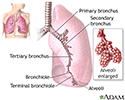
Lungs - illustration
The major features of the lungs include the bronchi, the bronchioles and the alveoli. The alveoli are the microscopic blood vessel-lined sacks in which oxygen and carbon dioxide gas are exchanged.
Lungs
illustration
Erythema multiforme, circular lesions - hands - illustration
Erythema multiforme lesions are circular and may appear in concentric rings (often called target lesions). Target lesions may also be associated with other medical conditions such as herpes infection, streptococcal infection, tuberculosis (TB), or as a reaction to chemicals or medications.
Erythema multiforme, circular lesions - hands
illustration
Erythema multiforme, target lesions on the palm - illustration
Erythema multiforme lesions are often referred to as target lesions because of the concentric rings the lesions produce. The target appearance is well demonstrated in this photograph.
Erythema multiforme, target lesions on the palm
illustration
Erythema multiforme on the leg - illustration
The red spots on this person's back appear where blisters (bullae) caused by Erythema multiforme have ruptured and the overlying skin removed (denuded). The resulting lesions are yellow-crusted ulcers (erosions). Erythema multiforme may be associated with herpes simplex infection, mycoplasma pneumonia, or other medical conditions such as streptococcal infection, tuberculosis (TB), or may result from exposure to chemicals or medications.
Erythema multiforme on the leg
illustration
Exfoliation following erythroderma - illustration
This picture shows diffuse redness (erythema) and scaling on the arm.
Exfoliation following erythroderma
illustration
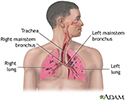
Respiratory system - illustration
Air is breathed in through the nasal passageways, travels through the trachea and bronchi to the lungs.
Respiratory system
illustration


Lungs - illustration
The major features of the lungs include the bronchi, the bronchioles and the alveoli. The alveoli are the microscopic blood vessel-lined sacks in which oxygen and carbon dioxide gas are exchanged.
Lungs
illustration
Erythema multiforme, circular lesions - hands - illustration
Erythema multiforme lesions are circular and may appear in concentric rings (often called target lesions). Target lesions may also be associated with other medical conditions such as herpes infection, streptococcal infection, tuberculosis (TB), or as a reaction to chemicals or medications.
Erythema multiforme, circular lesions - hands
illustration
Erythema multiforme, target lesions on the palm - illustration
Erythema multiforme lesions are often referred to as target lesions because of the concentric rings the lesions produce. The target appearance is well demonstrated in this photograph.
Erythema multiforme, target lesions on the palm
illustration
Erythema multiforme on the leg - illustration
The red spots on this person's back appear where blisters (bullae) caused by Erythema multiforme have ruptured and the overlying skin removed (denuded). The resulting lesions are yellow-crusted ulcers (erosions). Erythema multiforme may be associated with herpes simplex infection, mycoplasma pneumonia, or other medical conditions such as streptococcal infection, tuberculosis (TB), or may result from exposure to chemicals or medications.
Erythema multiforme on the leg
illustration
Exfoliation following erythroderma - illustration
This picture shows diffuse redness (erythema) and scaling on the arm.
Exfoliation following erythroderma
illustration
Respiratory system - illustration
Air is breathed in through the nasal passageways, travels through the trachea and bronchi to the lungs.
Respiratory system
illustration
Review Date: 8/19/2024
Reviewed By: Allen J. Blaivas, DO, Division of Pulmonary, Critical Care, and Sleep Medicine, VA New Jersey Health Care System, Clinical Assistant Professor, Rutgers New Jersey Medical School, East Orange, NJ. Review provided by VeriMed Healthcare Network. Also reviewed by David C. Dugdale, MD, Medical Director, Brenda Conaway, Editorial Director, and the A.D.A.M. Editorial team.
 All rights reserved.